Tuesday, December 19, 2006
Converting between Mci, mgRaeq and Air Kerma
How does one convert between Mci, mgRaeq and Air Kerma for different radioisotopes?
Note on Compton Scattering
In Compton scattering High Energy Photons (10 to 100 Mev) give most of their energy to the Compton electron, while low energy photons give most of their energy to the scattered photon.
Bragg-Gray vs Spencer Attix
Discuss Bragg-Gray theory and how this differs from Spencer-Attix theory.
DRR writes:
Before discussing the actual theory, I'll note that a big difference is that the Spencer Attix formulation accounts for the effects of secondary electrons produced.
DRR writes:
Before discussing the actual theory, I'll note that a big difference is that the Spencer Attix formulation accounts for the effects of secondary electrons produced.
Wednesday, December 13, 2006
Stopping Power
Discuss the difference between Stopping Power and Restricted Stopping Power.
Podgorsak pg 25.
The stopping power focuses on the energy loss by an electron moving
through a medium. When attention is focused on the absorbing medium,
one is interested in the linear rate of energy absorption by the absorbing
medium as the electron traverses the medium. The rate of energy
absorption, called the linear energy transfer (LET), is defined as the
average energy locally imparted to the absorbing medium by an electron
of specified energy in traversing a given distance in the medium.
● In radiation dosimetry the concept of restricted stopping power (SD/r) is
introduced, which accounts for that fraction of the collisional stopping
power (S/r)col that includes all the soft collisions plus those hard collisions
that result in delta rays with energies less than a cut-off value D. In
radiation dosimetry this cut-off energy is usually taken as 10 keV, an
energy that allows an electron just to traverse an ionization chamber gap
of 1 mm in air. Delta rays are defined as electrons that acquire sufficiently
high kinetic energies through hard collisions so as to enable them to carry
this energy a significant distance away from the track of the primary
particle and produce their own ionizations of absorber atoms.
TSET
QUESTION: Shown a diagram depicting a 6 field total skin electron irradiation. Explain the benefits of
such a setup. Be prepared to discuss clinical specifics (e.g. dose prescription, fractionation pattern, dose
inhomogeneity, energy, energy degraders, photon contamination, any required special physics
measurements).
such a setup. Be prepared to discuss clinical specifics (e.g. dose prescription, fractionation pattern, dose
inhomogeneity, energy, energy degraders, photon contamination, any required special physics
measurements).
Monday, December 11, 2006
Sunday, December 10, 2006
Saturday, December 9, 2006
Bragg Peak
What is the primary reason we see a Bragg Peak for heavy particles but not for electrons?
Wednesday, December 6, 2006
Designing a cesium safe
How would you construct a safe to store 500 mCi of Cs-137. What dose restrictions would you use? What materials would you use to construct it?
Dose algorithms (KERNAL)
Explain the term "kernal" and describe algorithms for electron beam calculation
Shielding considerations for a linac with energy >10MV
What are the shielding considerations for a linac with energy >10MV?
Monday, December 4, 2006
Work Load, U
With regards to shielding and radiation protection, what are values for the work load with regard to different locations in a clinic?
Effective Dose Limits
What are the effective dose limits for a member of the general public? What are they for a radiation worker?
I-125 vs Pd-103
Why would one use I-125 vs Pd-103 for prostate? What are the differences between them.
Thursday, November 30, 2006
Exposure Rate Constant (expressed)
What are the units of Exposure Rate constant?
Exposure Rate Constant is expressed in terms of R.cm2/hr.mCi
Exposure Rate Constant is expressed in terms of R.cm2/hr.mCi
Wednesday, November 29, 2006
Tuesday, November 28, 2006
Window and Level
Oral Question 2002 pg 233
Examinee was shown two pictures and asked what the difference was between them. They were the same CT slice with different window and levelling.
DEFINE WINDOW AND LEVEL
Examinee was shown two pictures and asked what the difference was between them. They were the same CT slice with different window and levelling.
DEFINE WINDOW AND LEVEL
Major components of a linac
What are the major components of a linac? Describe each of them and how they operate.
ACCELERATOR STRUCTURE which is either a Traveling wave or a standing wave.
Traveling Wave- High freq microwaves of 3000 MHz are transmitted down an evacuated tube through evenly spaced accelerating cavities (Lambda/4 which is approximately 2.5 cm) in length
A prebuncher is used to reduce velocity of the electromagnetic wave in order to correspond to the speed of the injected electron, so that the electron remains on the crest of the wave ("surfing") and undergoes acceleration. Electromagnetic waves are absorbed in a dummy load at the end of the guide to prevent them from reflecting and interfering with incoming waves.
Standing Wave- Standing wave is produced when two traveling waves of equal amplitude and period travel through a waveguide in opposite directions. Standing wave accelerator structures are used in most modern LINACS.
Electron Gun- Cathode that provides a source of bunched or pulsed electrons injected into an accelerator structure
Magnetron- "Makes Microwaves" . Produces them with frequency of 3000 MHz
Klystron- Carries Microwaves. It's a microwave amplifier driven by a low power microwave oscillator.
Modulator- Simulatenously produces high voltage direct current pulses to the magnetron or klystron and the electron gun.
Waveguide- Carries microwave power from a magnetron or klystron through the accelerator structure.
Accelerator Structure- Accelerates electrons from an electron gun using microwave power from a magnetron or klystron.
Treatment Head (Bending Magnet, Scattering foil, Target, Flattening Filter)
Add Schematic
Wednesday, November 8, 2006
Tandem and ovoid quick facts
DRR writes:
Location of point A and point B
Point A is located 2 cm laterally from the uterine canal and 2 cm above the lateral fornix. Point B is located on the pelvic wall 3 cm lateral to point A. The total dose to points A and B is the sum of the contributions from each source. WMK writes: It's a little more complicated than this. I'll comment later.
Sunday, October 22, 2006
Magnetron vs klystron
What are the differences. What are advantages/disadvantages of each?
Magnetron: Produces microwaves with v=3000 MHz, orginally used for radar and microwave ovens, operates at about 2 MW peak output, typically used to produce electron beams up to 15 MeV. MAGNETRON MAKES MICROWAVES
Klystron: Microwave amplifier driven by a low power microwave oscillator, operates at a peak output of 5 MW. KLYSTRON "Carries" MICROWAVES.
KLYSTRON is perceived to be more durable but is more expensive.
Thyratron
DRR writes:
Hendee (Radiation Therapy Physics p 62) states that the thyratron is a switching tube and is essentially a gas filled triode. When the grid is charged positively, the electrons flow from the cathode to the anode. The gas within the tube ionizes and conducts larger currents than do other switching devices. At the end of the pulse, the grid voltage is removed, preventing further current flow while the pulse forming network recharges. This cycle is repeated between 50 and 500 times per second.
Hendee (Radiation Therapy Physics p 62) states that the thyratron is a switching tube and is essentially a gas filled triode. When the grid is charged positively, the electrons flow from the cathode to the anode. The gas within the tube ionizes and conducts larger currents than do other switching devices. At the end of the pulse, the grid voltage is removed, preventing further current flow while the pulse forming network recharges. This cycle is repeated between 50 and 500 times per second.
Monday, October 9, 2006
MOSFETS
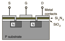
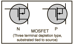
MOSFET (metal- oxide semiconductor field effect transistor)
MOSFET 3-Terminal
The Metal Oxide Semiconductor Field Effect Transistor (MOSFET) builds upon the basic FET by adding a layer of silicon dioxide as illustrated schematically. It achieves extremely high input impedance, in the range 109 to 1014 ohms.
Virtual SSD
Describe the steps necessary for determining the virtual SSD of a linac. Why would we want to perform this?
Wednesday, September 13, 2006
Uveal Melanoma
Uveal melanoma
Treatment-Typically 80 Gy to the apex of the tumor. I-125 seeds applied in an eye plaque applicator. Number of seeds can range (on the order of 10-15 seeds with average activity can vary greatly but from 2-8 mCi per seed which is high compared to the average seed used for prostate brachytherapy)
Malignant melanoma, the most common primary intraocular malignancy, is a neoplasm of the uveal tract. This is the pigmented layer of the eye that includes the iris, ciliary body, and choroid. The iris is the readily visible, most anterior portion. While the iris is perceived as giving the eye its color, such as blue, green, hazel, or brown, the melanocyte is the only pigment-synthesizing cell of the uveal tract. The amount of melanin varies according to racial and familial characteristics, and light diffraction explains the other aspects of iris color. The iris functions as a diaphragm, constantly altering the size of the pupil according to the ambient light.
The ciliary body is continuous with the iris, and it lines the sclera anteriorly. Its functions include secretion of aqueous fluid and alteration of the shape of the crystalline lens for the purpose of focusing. Posterior to it is the choroid, which lines the remainder of the sclera and functions as a source of oxygen and nutrients for the overlying retina. (https://www.moffitt.usf.edu/pubs/ccj/v5n4/article1.html)
One of the best description of this disease comes courtesy of this site:
http://www.medcyclopaedia.com/library/topics/volume_vi_1/u/uveal_melanoma.aspx
A more disturbing look at causes of uveal melanomas is the following (courtesy of http://www.biomedcentral.com/1471-2415/4/11)
Until now, the majority of epidemiological cancer studies focussed on brain cancer because the brain may be exposed to RFR [18-20]. With the exception of one study by Hardell et al. [21], all brain cancer studies showed no association between RFR as emitted by mobile phones and brain tumour risk until now. In contrast, the pooled analysis of two recent German case-control studies on the aetiology of uveal melanoma showed that frequent use of radiofrequency radiation devices including radio sets and mobile phones at the work place is associated with an about 4.2-fold elevated risk for uveal melanoma [22].
Saturday, August 19, 2006
What is the function of a beam spoiler?
Beam spoiler vs bolus, when would you use each.
DRR writes
A beam spoiler may be a low atomic number absorber, such as a Lucite shadow tray which is placed at an appropriate distance from the surface and is utilized to modify the buildup curve (Khan pg 281)
While a graph would represent it best and I should add one, basically a beam spoiler will increase the buildup at the surface while shifting the point of maximum dose buildup closer to the surface.
Saturday, July 15, 2006
Energy and PE, Compton and Pair Production
DRR (from conversation with WMK)
For interactions in water
At 25 keV, we have approximately 50% of events following Photoelectric effect and 50% events by Compton effect
At 150 keV, we have 100% Compton Effect
At 25 MeV we have 50% Compton, 50% Pair Production
For interactions in water
At 25 keV, we have approximately 50% of events following Photoelectric effect and 50% events by Compton effect
At 150 keV, we have 100% Compton Effect
At 25 MeV we have 50% Compton, 50% Pair Production
Tuesday, July 11, 2006
Friday, June 2, 2006
Arteriovenous Malformations
WMK:
An Arteriovenous Malformation, or AVM, is an abnormal collection of blood vessels. Normally, oxygenated blood is pumped by arteries to the brain, where it enters a fine network of tiny capillaries. It is in these capillary beds where the blood nourishes the tissues. The deoxygenated blood then passes back to the heart through veins. Arteriovenous malformations are areas that lack the tiny capillaries. The location of the connection between the artery and the vein is called the shunt. The area of tissue is called a nidus of the AVM. An AVM can be thought of as a "Short Circuit" where the blood does not go to the tissues but is pumped through the shunt and back to the heart without ever giving nutrients to the tissues.
What causes most AVMs is not known. People are born with AVMs although they do not appear to be hereditary. AVMs occur about equally in both sexes and in different races. An estimation of 3 million people in the United States are born with vascular malformation, 10% of which are AVMs.
Most patients do not know that they have an AVM. A number of the patients with AVMs have seizures or persistent headaches. An AVM can put additional strain on the blood vessels and the surrounding tissues. For the very young (under the age of twenty) this is usually not a problem. The increased flow of blood caused by the shunt weakens the blood vessels. These weakened blood vessels can rupture. This is known as a hemorrhage or a bleed. If an AVM bleeds, the patient experiences an extremely severe headache. The bleed may cause a stroke and even death. About 4% of people with AVMs experience initial bleeds each year.
AVMs can be seen on outpatient imaging studies such as CT's or MRI's. Angiograms are inpatient procedures needed to image the arteries and veins before any treatment. An angiogram is an x-ray movie of the blood flowing through the blood vessels. It is made by injecting contrast into the arteries going into the head and taking a series of x-rays films.
Treatment options for AVMs include embolization, radiation, and surgery or a combination of these methods. Recent studies have revealed that for most cases, embolization is the safest and most effective procedure. To avoid bleeding, the aneurysm must be eliminated. Each treatment has its advantages and disadvantages.
Embolization is a method of plugging the blood vessels of the AVM. Under X-ray guidance, a catheter is guided from the femoral artery in the leg up into the area to be treated. Once the area is reached, glue or sometimes even a wire coil is placed to block off the area.
Electron Blocks
WMK:
We read an electron block if the size of the opening in centimeters is less than half the energy in MeV. For example, for a 9 MeV block, if the opening is less than 4.5 cm. near the center of the field we must make a physical measurement. If the opening is greater than 4.5 cm. we can make a calculation as if the block was open.
When measuring a block, we make two readings. One reading is where we know the output at and the other is at the treatment distance. For example, we can take an open field measurement at 100 cm. For a 10 x 10 field at 100 cm. the output is 1 cGy/MU at dmax. We record the electrometer reading. This gives us the reading to deliver 1 cGy/MU. Next we take a reading for the blocked field at the treatment distance. This reading divided by the open field known reading will give us the cGy/MU delivered under the treatment conditions.
We use this information to calculate the monitor units necessary to deliver the prescribed dose.
HRE adds these comments:
We're making a few assumptions here. One is that fractional depth dose doesn't change between your open measurement and your treatment set up measurement. That is, the distance of dmax remains the same for the two measurements. Another assumption is that the electrometer reading is linearly proportional to dose.
DRR adds this:
Note that for purposes of the calculation, we use the electron cone factor from the table at 100 SSD if we measured the open field at this distance. A common mistake when a treatment is at 110 SSD and the block is read would be to use the electron cone factor at 110 SSD. This would be wrong.
Subscribe to:
Posts (Atom)